FUNDUS PHOTOGRAPHY
What Is Fundus Photography?
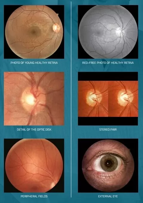
Fundus photography is taken by a specialist fundus camera. It has a low-powered microscope with a camera attachment which is able to capture a detailed photograph of the back of your eye.It photographically captures detail of the retina. optic disc, macula and posterior pole (fundus).
Fundus photography is used for diagnosing disease and monitoring any progression of that disease over time.
WHY DO I NEED A FUNDUS PHOTOGRAPH TAKEN OF MY EYES?
A fundus photograph can detect and monitor serious conditions such as; Glaucoma, Diabetes, Age-related Macula Degeneration (AMD) and other conditions such as Papilledema.
HOW LONG WILL THE PROCEDURE TAKE?
The procedure is very quick, and it will only take around 1 minute to take a photograph of both eyes.
WHAT HAPPENS WHEN I HAVE THE PHOTOGRAPHS TAKEN?
Our optometrist will ask you to place your chin on a chin rest and your forehead against a support bar. You will then be asked to stare at a green light, which will help you to look into the correct position.
You will see a series of flashes while the photos are taken. The test is non-contact and there are no sudden puffs of air.
WHAT HAPPENS NEXT?
Our optometrist will look carefully at the photographs to check for signs of any potential health issues. We will recommend that you have a photograph taken at each future appointment, in order to record and monitor any changes in your eye.
If there is a cause for concern, your photographs can be used to refer you to a specialist.
Read our Google reviews from delighted patients and customers below - and why not add your own?
Opening Hours
Monday 9am-5:30pm
Tuesday 9am-5:30pm
Wednesday 9am-5:30pm
Thursday 9am-5:30pm
Friday 9am-5:30pm
Saturday 9am-5pm
Sunday CLOSED
Designer Eyewear
Contact Us
Connect With Us